Performance and Analysis of a U-Net Model for Automated Skin Lesion Segmentation
Article Sidebar
Main Article Content
Abstract
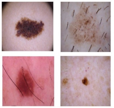
A greater proportion of people are affected by skin cancer, particularly melanoma, which has a higher tendency to metastasize. For Dermatologist, Visual inspections are most challenging & complex task for melanoma detection. To solve this problem, dermoscopic images are analyzed and segmented. Due to the sensitivity involved in surgical operations, existing techniques are unable to achieve higher accuracy. As a result, computer-aided systems are essential to detect & segment dermoscopic images.
In this paper, for segmentation 5000 skin images were taken from the HAM10000 dataset. Prior to segmentation, preprocessing is done by resizing images. A novel U Net structure is a fully convolutional network is presented & implemented using up-sampling and down-sampling technique with Rectified Linear Units (ReLU) for activation functions. The outcomes of proposed methodology shows performance improvement for skin-lesion segmentation with 94.7 % pixel accuracy & 89.2 % dice coefficient compared with existing KNN & SVM techniques.
Article Details
References
Iqbal, A.; Sharif, M.; Yasmin, M.; Raza, M.; Aftab, S, “Generative adversarial networks and its applications in the biomedical image segmentation: A comprehensive survey”, Int. J. Multimed. Inf. Retr, pp. 333–368, Nov 2022
Masood, S.; Sharif, M.; Masood, A.; Yasmin, M., “Raza, M. A Survey on Medical Image Segmentation”, Curr. Med. Imaging, pp. 3-14, Nov 2015
Anjum, M.A.; Amin, J.; Sharif, M.; Khan, H.U.; Malik, M.S.A.; Kadry, S., “Deep Semantic Segmentation and Multi-Class Skin Lesion Classification Based on Convolutional Neural Network”, IEEE Access, pp. 129668–129678, Aug 2020
Sayel M. Fayyad, Mohammad Abuzalatah, Mohannad Rawashdeh, A. M. Maqableh, Zaid Abulghanam. (2023). Control, Design and Analysis of Delta 3D Printer . International Journal of Intelligent Systems and Applications in Engineering, 11(4s), 444–457. Retrieved from https://ijisae.org/index.php/IJISAE/article/view/2702
Iqbal, A., Sharif, M., Khan, M.A. et al, “FF-UNet: a U-Shaped Deep Convolutional Neural Network for Multimodal Biomedical Image Segmentation”, Cogn Comput 14, 1287–1302 (2022).
Shahzad, A.; Sharif, M.; Raza, M.; Hussain, K., “Enhanced watershed image processing segmentation”, J. Inf. Commun. Technol. Vol. 2, No. 1, pp.01-09, Spring 2008.
Na Hwang, Y.; Seo, M.J.; Kim, S.M, “A Segmentation of Melanocytic Skin Lesions in Dermoscopic and Standard Images Using a Hybrid Two-Stage Approach”, BioMed Res. Int. 2021,April 2021, 5562801
Garg, S.; Jindal, B, “Skin lesion segmentation using k-mean and optimized fire fly algorithm”, Multimed. Tools, pp.7397–7410, Appl. 2021, 80.
Hawas, A.R.; Guo, Y.; Du, C.; Polat, K.; Ashour, A.S, “OCE-NGC: A neutrosophic graph cut algorithm using optimized clustering estimation algorithm for dermoscopic skin lesion segmentation”, Appl. Soft Comput. 2020, 86, 105931
Dr. S.A. Sivakumar. (2019). Hybrid Design and RF Planning for 4G networks using Cell Prioritization Scheme. International Journal of New Practices in Management and Engineering, 8(02), 08 - 15. https://doi.org/10.17762/ijnpme.v8i02.76
Mohamed, A.A.I.; Ali, M.M.; Nusrat, K.; Rahebi, J.; Sayiner, A.; Kandemirli, F, “Melanoma skin cancer segmentation with image region growing based on fuzzy clustering mean”, Int. J. Eng. Innov. Res. 2017, 6, 91C95.
Jaisakthi, S.M.; Chandrabose, A.; Mirunalini, P, “Automatic skin lesion segmentation using semi-supervised learning technique”, arXiv 2017, arXiv:1703.04301.
Lynn, N.C.; Kyu, Z.M, “Segmentation and Classification of Skin Cancer Melanoma from Skin Lesion Images”, In Proceedings of the 2017 18th International Conference on Parallel and Distributed Computing, Applications and Technologies (PDCAT), Taipei,Taiwan, pp. 117–122,18–20, December 2017.
Saravanan, S.; Heshma, B.; Shanofer, A.A.; Vanithamani, R, “Skin cancer detection using dermoscope images”, Mater. Today: , pp. 4823–4827,Proc.2020, 33.
Thanh, D.N.H.; Erkan, U.; Prasath, V.S.; Kumar, V.; Hien, N.N, “A Skin Lesion Segmentation Method for Dermoscopic Images Based on Adaptive Thresholding with Normalization of Color Models”, In Proceedings of the 2019 6th International Conference on Electrical and Electronics Engineering (ICEEE), Istanbul, Turkey, 16–17 April 2019.
Abdulhamid, I.A.M.; Sahiner, A.; Rahebi, J, “New Auxiliary Function with Properties in Nonsmooth Global Optimization for Melanoma Skin Cancer Segmentation”, BioMed Res. Int. 2020, 2020, 5345923
Javed, R.; Rahim, M.S.M.; Saba, T.; Rashid, M, “Region-based active contour JSEG fusion technique for skin lesion segmentation from dermoscopic images”, pp.1–10, Biomed. Res. 2019, 30.
Ashour, A.S.; Nagieb, R.M.; El-Khobby, H.A.; Elnaby, M.M.A.; Dey, N, “Genetic algorithm-based initial contour optimization for skin lesion border detection”, Multimed. Tools, pp. 2583–2597 Appl. 2021, 80.
Mohakud, R.; Dash, R, “Skin cancer image segmentation utilizing a novel EN-GWO based hyper-parameter optimized FCEDN”, J. King Saud Univ.-Comput. Inf. Sci, pp. 9889–9904, 2022, 34.
Kaur, R.; Gholam, H.; Sinha, R.; Lindén, M, “Automatic lesion segmentation using atrous convolutional deep neural networks in dermoscopic skin cancer images”, BMC Med. Imaging, pp.1–13, 2021, 22.
Bagheri, F.; Tarokh, M.J.; Ziaratban, M, “Skin lesion segmentation from dermoscopic images by using Mask R-CNN, Retina-Deeplab,and graph-based methods”, Biomed. Signal Process. Control. 2021, 67, 102533.
Emma Smith, Deep Learning for Gesture Recognition and Human-Computer Interaction , Machine Learning Applications Conference Proceedings, Vol 3 2023.
Qamar, S.; Ahmad, P.; Shen, L, “Dense Encoder-Decoder–Based Architecture for Skin Lesion Segmentation”, Cogn. Comput, pp.583–594, 2021,13.
Wu, H.; Pan, J.; Li, Z.; Wen, Z.; Qin, J, “Automated Skin Lesion Segmentation Via an Adaptive Dual Attention Module”, IEEE Trans.Med. Imaging, pp.357–370, 2020, 40.
Öztürk, ¸S.; Özkaya, U, “Skin Lesion Segmentation with Improved Convolutional Neural Network”. J. Digit. Imaging, pp.958–970, 2020, 33.
Nezhadian, F. K. , and Rashidi, S, “ Melanoma skin cancer detection using color and new texture features” 2017 Artificial Intelligence and Signal Processing Conference (AISP), Shiraz. pp.1–5, 2017.
Mustafa, S. and Kimura, A, “A SVM-based diagnosis of melanomausing only useful image features”, 2018 International Workshop onAdvanced Image Technology (IWAIT), ChiangMai. pp.1–4, 2018.
Waheed, Z., Waheed, A., Zafar, M. and Riaz, F, “ An efficient machinelearning approach for the detection of melanoma usingdermoscopic images”, 2017 International Conference onCommunication, Computing and Digital Systems (C-CODE),Islamabad. pp.316–319, 2017.
Silveira, M., Nascimento, J. C., Marques, J. S., Marçal, A. R.,Mendonça, T., Yamauchi, S., Maeda, J., and Rozeira, J,“Comparison of segmentation methods for melanoma diagnosis in dermoscopy images”, IEEE J. Sel. Top. Signal Process 3(1):pp.35–45,2009
Pennisi, A., Bloisi, D.D.,Nardi, D., Giampetruzzi, A. R.,Mondino,C., and Facchiano, A, “ Skin lesion image segmentation using Delaunay triangulation for melanoma detection” Comput. Med. Imaging Graph. 52:89–103, 2016.
Maglogiannis, I., Doukas, C. N., and Member, S, “Overview ofadvanced computer vision Systems for Skin LesionsCharacterization”, IEEE Trans. Inf. Technol. Biomed.13(September):721–733, 2009.
Tushar H. Jaware , K. B. Khanchandani , Anita Zurani, “An Accurate Automated Local Similarity Factor-Based Neural Tree Approach toward Tissue Segmentation of Newborn Brain MRI ”, Am J Perinatol 2019; 36(11): 1157-1170, DOI: 10.1055/s-0038-1675375
Jaware, T.H., Patil, V.R., Badgujar, R.D. et al. Performance investigations of filtering methods for T1 and T2 weighted infant brain MR images. Microsyst Technol 27, 3711–3723 (2021). https://doi.org/10.1007/s00542-020-05144-6
Tushar Jaware,Kamlesh Khanchandani &Ravindra Badgujar, “A novel hybrid atlas-free hierarchical graph-based segmentation of newborn brain MRI using wavelet filter banks” International Journal of Neuroscience, vol 130 (5), pp 499–514, 2020
P. G. Patil et al., "Marathi Speech Intelligibility Enhancement Using I-AMS Based Neuro-Fuzzy Classifier Approach for Hearing Aid Users," in IEEE Access, vol. 10, pp. 123028-123042, 2022, doi: 10.1109/ACCESS.2022.3223365.