An Integrated Framework for the Detection of Lung Nodules from Multimodal Images Using Segmentation Network and Generative Adversarial Network Techniques
Article Sidebar
Main Article Content
Abstract
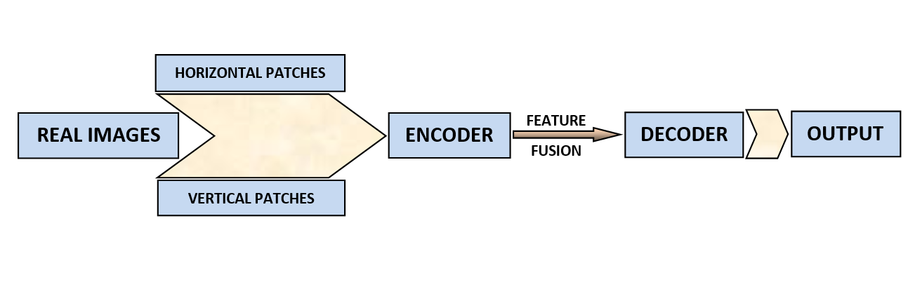
Medical imaging techniques are providing promising results in identifying abnormalities in tissues. The presence of such tissues leads to further investigation on these cells in particular. Lung cancer is seen widely and is deadliest in nature if not detected and treated at an early stage. Medical imaging techniques help to identify the presence of suspicious tissues like lung nodules effectively. But it is very difficult to know the presence of the nodule at an early stage with the help of a single imaging modality. The proposed system increases the efficiency of the system and helps to identify the presence of lung nodules at an early stage. This is achieved by combining different methods for reaching a common outcome. Multiple schemes are combined and the extracted features are used for obtaining a conclusion. The accuracy of the system and the results depend on the quality and quantity of the authentic training data. But the availability of the data from an authentic source for the study is a challenging task. Here the generative adversarial network (GAN), is used as a data source generator. It helps to generate a huge amount of reliable data by using a minimum number of real time and authentic data set. Images generated by the GAN are of resolution 1024 x 1024.Fine tuning of the images by using the real images increases the quality of the generated images and thereby improving the efficiency. Luna 16 is the primary data source and these images are used for the generation of 1000000 images. Training process with the huge dataset improves the capability of the proposed system. Various parameters are considered for evaluating the performance of the proposed system. Comparative analysis with existing systems highlights the strengths of the proposed system.
Article Details
References
P. D. Mozley et al., “Measurement of Tumor Volumes Improves RECIST-Based Response Assessments in Advanced Lung Cancer,” Transl. Oncol., vol. 5, no. 1, pp. 19–25, Feb. 2012.
S. A. Hayes et al., “Comparison of CT volumetric measurement with RECIST response in patients with lung cancer,” Eur. J. Radiol., vol. 85, no. 3, pp. 524–533, Mar. 2016.
M. Chen, X. Shi, Y. Zhang, D. Wu, and M. Guizani, “Deep Features Learning for Medical Image Analysis with Convolutional Autoencoder Neural Network,” IEEE Trans. Big Data, 2017.
J. N. S. S., J. N. ., & E. N., G. . (2023). A Novel Blockchain-Based Lightweight Encryption Technique in Fog Based IoT for Personal Healthcare Data Application. International Journal of Intelligent Systems and Applications in Engineering, 11(3s), 119 –. Retrieved from https://ijisae.org/index.php/IJISAE/article/view/2549
W. Chen, H. Wei, S. Peng, J. Sun, X. Qiao, and B. Liu, “HSN: Hybrid Segmentation Network for Small Cell Lung Cancer Segmentation,” IEEE Access, vol. 7, pp. 75591–75603, 2019.
Y. Gordienko et al., “Deep Learning with Lung Segmentation and Bone Shadow Exclusion Techniques for Chest X-Ray Analysis of Lung Cancer,” 2019, pp. 638–647.
F. Isensee et al., “nnU-Net: Self-adapting Framework for U-Net-Based Medical Image Segmentation,” Sep. 2018.
H.-C. Shin et al., “Deep Convolutional Neural Networks for Computer-Aided Detection: CNN Architectures, Dataset Characteristics and Transfer Learning,” IEEE Trans. Med. Imaging, vol. 35, no. 5, pp. 1285–1298, May 2016.
P. Tschandl, C. Sinz, and H. Kittler, “Domain-specific classification-pretrained fully convolutional network encoders for skin lesion segmentation,” Comput. Biol. Med., vol. 104, pp. 111–116, Jan. 2019.
J. Gong, J. Y. Liu, X. W. Sun, B. Zheng, and S. D. Nie, “Computer-aided diagnosis of lung cancer: The effect of training data sets on classification accuracy of lung nodules,” Phys. Med. Biol., 2018.
H. Xie, D. Yang, N. Sun, Z. Chen, and Y. Zhang, “Automated pulmonary nodule detection in CT images using deep convolutional neural networks,” Pattern Recognit., vol. 85, pp. 109–119, 2019.
V. C and M. D, “Multimodal Data Analysis Using Soft Computing Techniques For The Detection and Classification of Lung Cancer,” in 2022 Third International Conference on Intelligent Computing Instrumentation and Control Technologies (ICICICT), 2022, pp. 150–153.
C. H. Chen et al., “Radiomic features analysis in computed tomography images of lung nodule classification,” PLoS One, 2018.
G. Litjens et al., “A survey on deep learning in medical image analysis,” vol. 42, no. December 2012, pp. 60–88, 2017.
Natalia Volkova, Machine Learning Approaches for Stock Market Prediction , Machine Learning Applications Conference Proceedings, Vol 2 2022.
S. Sasikala, M. Bharathi, and B. R. Sowmiya, “Lung cancer detection and classification using deep CNN,” Int. J. Innov. Technol. Explor. Eng., vol. 8, no. 2S, pp. 259–262, 2018.
A. A. Farag, A. Ali, S. Elshazly, and A. A. Farag, “Feature fusion for lung nodule classification,” Int. J. Comput. Assist. Radiol. Surg., 2017.
M. Nishio et al., “Attribute-guided image generation of three-dimensional computed tomography images of lung nodules using a generative adversarial network,” Comput. Biol. Med., vol. 126, p. 104032, Nov. 2020.
H. Cao et al., “A Two-Stage Convolutional Neural Networks for Lung Nodule Detection,” IEEE J. Biomed. Heal. Informatics, 2020.
W. Li, P. Cao, D. Zhao, and J. Wang, “Pulmonary Nodule Classification with Deep Convolutional Neural Networks on Computed Tomography Images,” vol. 2016, 2016.
H. Cao et al., “Dual-branch residual network for lung nodule segmentation,” Appl. Soft Comput. J., 2020.
M. Nishio, S. Noguchi, H. Matsuo, and T. Murakami, “Automatic classification between COVID-19 pneumonia, non-COVID-19 pneumonia, and the healthy on chest X-ray image: combination of data augmentation methods,” Sci. Rep., vol. 10, no. 1, p. 17532, Dec. 2020.
K. Armanious et al., “MedGAN: Medical image translation using GANs,” Comput. Med. Imaging Graph., vol. 79, p. 101684, Jan. 2020.
Schmid-Bindert G, Vogel-Claussen J, Gütz S, Fink J, Hoffmann H, Eichhorn ME, Herth FJF. Incidental Pulmonary Nodules - What Do We Know in 2022. Respiration. 2022;101(11):1024-1034. doi: 10.1159/000526818. Epub 2022 Oct 13. PMID: 36228594; PMCID: PMC9945197
Wang H, Zhu H, Ding L. Accurate classification of lung nodules on CT images using the TransUnet. Front Public Health. 2022 Dec 5;10:1060798. doi: 10.3389/fpubh.2022.1060798. PMID: 36544802; PMCID: PMC9760709.