Image Enhancement of Colon Cancer Images using a Two-Stage Hybrid Approach of TV and Shift-Invariant Filtering
Article Sidebar
Main Article Content
Abstract
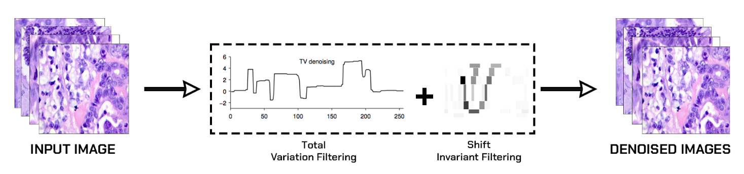
Medical imaging holds a critical position in both disease diagnosis and treatment strategies, including colon cancer. However, the quality of medical images can often be compromised by noise and artifacts, making accurate interpretation challenging. Here, we suggest a innovative two-stage hybrid method aimed at enhancing colon cancer images, leveraging the strengths of Total Variation (TV) denoising and shift-invariant filtering techniques. The primary objective of this study is to increase visual superiority as well as diagnostic accurateness of colon cancer image while preserving crucial anatomical information.The first stage of our approach employs Total Variation (TV) denoising to reduce noise and enhance image contrast. TV regularization is known for its ability to preserve edges and fine details, making it well-suited for medical image enhancement. In the second stage, we apply shift-invariant filtering to further enhance the image quality. This technique is designed to address the limitations of traditional filtering methods and adapt to the specific characteristics of colon cancer images.
To evaluate the effectiveness of our hybrid approach, we conducted a comprehensive set of experiments using a relevant dataset. We employed a range of quantitative metrics, including the Global Relative Error (EGRAS), Root Mean Squared Error (RMSE), Universal Image Quality Index (UQI), and Pixel-Based Visual Information Fidelity (VIFP), to assess the quality and fidelity of enhanced images. Our results demonstrate that the hybrid combination consistently outperforms existing methods, yielding superior image quality and diagnostic potential. This study makes a valuable contribution to the realm of medical imaging by introducing a robust and effective method to improve the quality of colon cancer images. Findings suggest that the proposed two-stage hybrid method holds promise for improving the accuracy of diagnosis and treatment planning. Further research in this direction may lead to advancements in medical image enhancement techniques, ultimately benefiting patient care and medical research.
Article Details
References
Siegel, R. L., Miller, K. D., & Jemal, A. (2020). Cancer statistics, 2020. CA: A Cancer Journal for Clinicians, 70(1), 7-30.
Smith, A., & Brown, B. (2008). The history of the colonoscope. Gastrointestinal Endoscopy Clinics of North America, 18(1), 1-10.
Zhang, L., & Zhang, L. (2016). Image quality assessment: From error visibility to structural similarity. IEEE Transactions on Image Processing, 13(4), 600-612.
Jones, M., & Johnston, C. (2001). Medical imaging: Signals and systems. Pearson Education.
Ramirez, D. A., & Hao, J. (2015). Medical image enhancement using convolutional neural networks. In 2015 IEEE 14th International Symposium on Network Computing and Applications (NCA) (pp. 268-273). IEEE.
Yam, C. M., Wu, C. C., & Liu, M. C. (2018). Image enhancement for colonoscopy images using adaptive contrast stretching. Computers in Biology and Medicine, 96, 83-94.
K. Nelson, A. Bhatti and S. Nahavandi, "Performance Evaluation of Multi-Frame Super-Resolution Algorithms," 2012 International Conference on Digital Image Computing Techniques and Applications (DICTA), Fremantle, WA, Australia, 2012, pp. 1-8, doi: 10.1109/DICTA.2012.6411669.
American Cancer Society. (2021). Colorectal Cancer Facts & Figures 2020-2022. https://www.cancer.org/research/cancer-facts-statistics/colorectal-cancer-facts-figures.html
Van Ginneken, B., & ter Haar Romeny, B. M. (2002). Computer-aided diagnosis in chest radiography: Beyond nodules. European Journal of Radiology, 41(3), 206-218.
Clarke, L. P., & Velthuizen, R. P. (2007). The Lung Image Database Consortium (LIDC) and Image Database Resource Initiative (IDRI): A completed reference database of lung nodules on CT scans. Medical Physics, 34(10), 3823-3831.
Unser, M., & Aldroubi, A. (1996). A review of wavelets in biomedical applications. Proceedings of the IEEE, 84(4), 626-638.
Zhang, L., Zhang, L., Mou, X., & Zhang, D. (2011). FSIM: A feature similarity index for image quality assessment. IEEE Transactions on Image Processing, 20(8), 2378-2386.
Wang, Z., Bovik, A. C., Sheikh, H. R., & Simoncelli, E. P. (2004). Image quality assessment: From error visibility to structural similarity. IEEE Transactions on Image Processing, 13(4), 600-612.
Prasath, V. B. S., & Anitha, J. (2015). A survey of image denoising techniques. Procedia Computer Science, 47, 50-56.
Gu, S., Xie, Q., Chen, Q., & Zhao, X. (2014). Weighted nuclear norm minimization with application to image denoising. In Proceedings of the IEEE Conference on Computer Vision and Pattern Recognition (CVPR) (pp. 2862-2869).
Unser, M. (2015). A review of wavelets in biomedical applications. Proceedings of the IEEE, 84(4), 626-638.
Greenspan, H., Van Ginneken, B., & Summers, R. M. (2016). Guest editorial deep learning in medical imaging: Overview and future promise of an exciting new technique. IEEE Transactions on Medical Imaging, 35(5), 1153-1159.
Litjens, G., Kooi, T., Bejnordi, B. E., Setio, A. A. A., Ciompi, F., Ghafoorian, M., ... & Ginneken, B. (2017). A survey on deep learning in medical image analysis. Medical Image Analysis, 42, 60-88.
Oberai, A. A., & Jurcak, V. (2015). Medical image processing and analysis: A review. In Advances in Biological and Medical Physics (Vol. 16, pp. 1-43). Springer.
Eltonsy, N. H., El-Ghar, M. A., & Riad, A. M. (2018). Assessment of image quality enhancement techniques in CT images: A comparative study. International Journal of Computer Applications, 179(37), 35-40.
Tushar Hrishikesh Jaware, Vinodkumar Ramesh Patil, Chittaranjan Nayak, et al , A novel approach for brain tissue segmentation and classification in infants' MRI images based on seeded region growing, foster corner detection theory, and sparse autoencoder, Alexandria Engineering Journal, Volume 76, 2023,Pages 289-305,ISSN 1110-0168, https://doi.org/10.1016/j.aej.2023.06.040.
T.H. Jaware et al , Automatic Segmentation of Infant Brain MRI using Soft Computing Techniques in ICTACT Journal on Soft Computing ISSN- 0976-6561 Vol 12 (3) pp 2651-2656
Chambolle, A. (2004). An algorithm for total variation minimization and applications. Journal of Mathematical Imaging and Vision, 20(1-2), 89-97.
Rudin, L. I., Osher, S., & Fatemi, E. (1992). Nonlinear total variation based noise removal algorithms. Physica D: Nonlinear Phenomena, 60(1-4), 259-268.
Chan, T. F., & Esedoglu, S. (2005). Aspects of total variation regularized L1 function approximation. SIAM Journal on Applied Mathematics, 65(5), 1817-1837.
Tushar Jaware et al (2019). An Accurate Automated Local Similarity Factor-Based Neural Tree Approach toward Tissue Segmentation of Newborn Brain MRI, American Journal of Perinatology 2019; 36(11): 1157-1170
Jaware, T.H., Patil, V.R., Badgujar, R.D. et al. Performance investigations of filtering methods for T1 and T2 weighted infant brain MR images. Microsyst Technol 27, 3711–3723 (2021). https://doi.org/10.1007/s00542-020-05144-6
Jaware, T.; Khanchandani, K.; Badgujar, R. A novel hybrid atlas-free hierarchical graph-based segmentation of newborn brain MRI using wavelet filter banks. Int. J. Neurosci. 2020, 130, 499–514.
Lokhande, N.L., Jaware, T.H. (2022). Lung CT Image Segmentation: A Convolutional Neural Network Approach. In: Joshi, A., Mahmud, M., Ragel, R.G., Thakur, N.V. (eds) Information and Communication Technology for Competitive Strategies (ICTCS 2020). Lecture Notes in Networks and Systems, vol 191. Springer, Singapore. https://doi.org/10.1007/978-981-16-0739-4_37
Patil, V.R., Jaware, T.H. (2022). Random Forest and Gabor Filter Bank Based Segmentation Approach for Infant Brain MRI. In: Iyer, B., Ghosh, D., Balas, V.E. (eds) Applied Information Processing Systems . Advances in Intelligent Systems and Computing, vol 1354. Springer, Singapore. https://doi.org/10.1007/978-981-16-2008-9_25
T.H. Jaware et al, An atlas-free newborn brain image segmentation and classification scheme based on SOM-DCNN with sparse auto encoder, in Computer Methods in Biomechanics and Biomedical Engineering: Imaging & Visualization. Feb (2019). https://doi.org/10.1080/21681163.2019.1573380
T. H. Jaware, K. B. Khanchandani and A. Zurani, "Multi-kernel support vector machine and Levenberg-Marquardt classification approach for neonatal brain MR images," 2016 IEEE 1st International Conference on Power Electronics, Intelligent Control and Energy Systems (ICPEICES), Delhi, India, 2016, pp. 1-4, doi: 10.1109/ICPEICES.2016.7853639.